Small objects, big stakes: detecting kidney stones at high resolution
The smallest target in the dataset sets the resolution budget for the whole model. Here is why Atlas reads CT at 1280 pixels and adds a high-resolution detection head.
The smallest target sets the budget
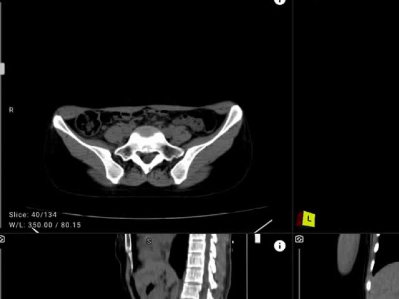
In our dataset, kidney stones have a median bounding box of about 39 pixels at native resolution, with the smallest 5 percent under 18 pixels. CT is commonly stored at 512 by 512 in-plane. At that size, a stone is a handful of pixels, and a detector that downsamples aggressively will simply lose it. The smallest clinically important object dictates the resolution the whole pipeline has to support.
1280 pixels and a stride-4 head
Atlas upsamples each slice to 1280 by 1280 and adds a stride-4 detection head, which produces a 320 by 320 feature map capable of localizing objects as small as about 8 pixels. That recovers recall on the smallest stones while preserving the fine soft-tissue detail that subtler findings, like fat stranding or wall thickening, depend on. Internally, kidney stones reached 81.7 percent F1.
Why a single-stage detector
Atlas uses a one-stage detector rather than a two-stage region-proposal network. Contemporary medical adaptations of single-stage detectors localize CT findings well [1], inference latency stays compatible with emergency triage, and the model footprint fits a hospital-grade GPU. Bounding-box regression uses Wise-IoU, which focuses learning on ordinary-quality boxes rather than chasing outliers [2].
The catch: contrast phase
Resolution is not the only variable for stones. Their conspicuity depends on contrast phase, because intravenous contrast can raise attenuation in ways that interact with the high-attenuation signature a stone detector relies on [3]. This is part of why kidney stones were one of the classes most sensitive to the move from the internal cohort to an external one [4], and why protocol-stratified evaluation is on the roadmap.
References
- Sobek J, et al. MedYOLO: a medical image object detection framework. J Imaging Inform Med. 2024;37(6):3208-3216.
- Tong Z, et al. Wise-IoU: bounding box regression loss with dynamic focusing mechanism. arXiv:2301.10051. 2023.
- Holcombe SA, et al. Variation in aorta attenuation in contrast-enhanced CT and implications for calcification thresholds. PLoS One. 2022;17(11):e0277111.
- Ye Y, et al. Kidney stone detection in CT using a YOLOv8 framework. Front Med. 2026;13:1702159.