EVREON AI · CLINICAL AGENT PLATFORM

Clinical AI agents for hospital imaging.

Purpose-built for emergency abdominal CT. Every finding localized, anatomy-checked, and ready for clinician review.

Project Atlas abdominal CT review interface showing a four-panel scan viewer, emergency analysis findings, and clinician review actions
PROJECT ATLASPilot-ready · Live capture

Evreon AI is the platform. Atlas is the first product.

Close-up view of a CT scanner in a hospital imaging room

PILOT READY

Project Atlas

Abdominal CT

Emergency abdominal CT agent for six urgent pathologies, localized findings, anatomy checks, and report drafts.

Active first product
Doctors reviewing brain MRI scans together

FUTURE

Project NeuroLens

Brain imaging

A future neuro-focused agent family for brain imaging workflows and anatomy-aware review.

Planned product line
Doctors reviewing a medical scan for future body-specific imaging agents

ROADMAP

Future body systems

Modality-specific agents

Each new Evreon product will keep its own clinical scope, model stack, and review workflow.
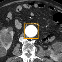
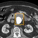
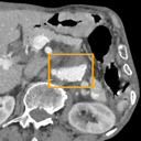
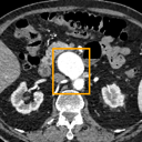
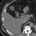
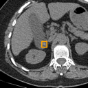
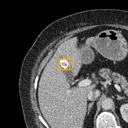
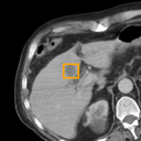
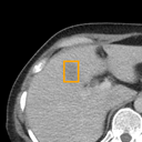
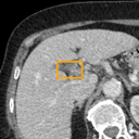
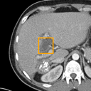
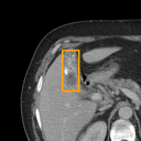
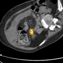
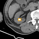
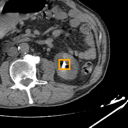
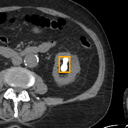
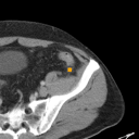
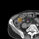
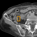
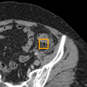
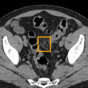
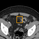
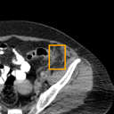
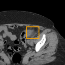
Platform direction

Agentic AI for abdominal CT.

Project Atlas analyzes abdominal CT scans for six urgent pathologies, localizes suspected findings in the scan, verifies them against anatomical structures, and prepares a draft clinical report for doctor review.

~85 million abdominal CT scans are performed each year across Europe and the US. Every one a time-critical read.

Six abdominal emergency pathologies

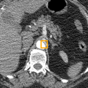
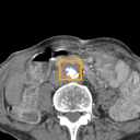
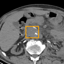
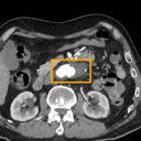
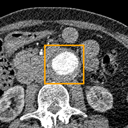
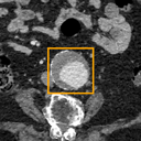
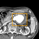
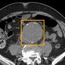
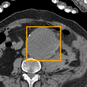
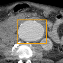
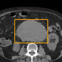
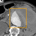
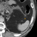
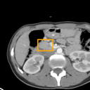
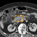
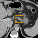
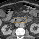
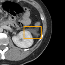
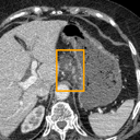
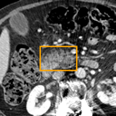
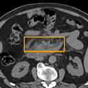
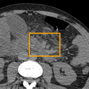
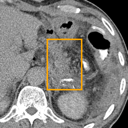
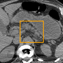
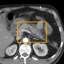
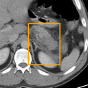
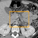
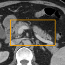
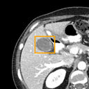
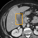
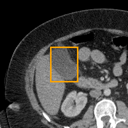
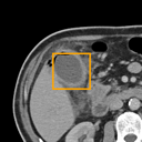
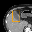
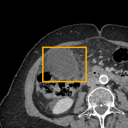
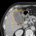
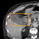
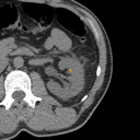
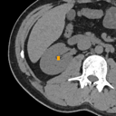
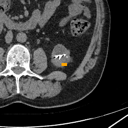
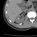
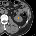
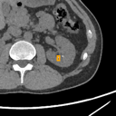
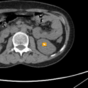
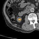
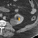
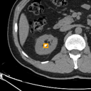
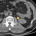
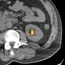
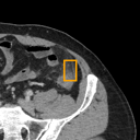
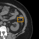
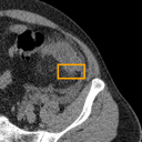
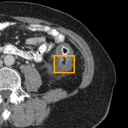
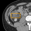
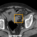
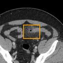
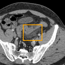
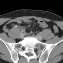
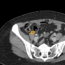
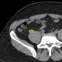
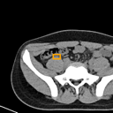
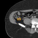
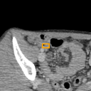
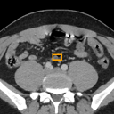
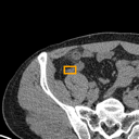
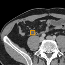
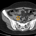
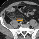
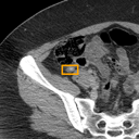
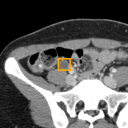
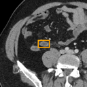
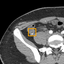
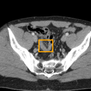
A deliberately locked clinical scope. Hover any card to inspect boxed reference views drawn from Atlas training data.

Abdominal Aortic Aneurysm bounding-box exemplar 1Abdominal Aortic Aneurysm bounding-box exemplar 2Abdominal Aortic Aneurysm bounding-box exemplar 3Abdominal Aortic Aneurysm bounding-box exemplar 4Abdominal Aortic Aneurysm bounding-box exemplar 5Abdominal Aortic Aneurysm bounding-box exemplar 6Abdominal Aortic Aneurysm bounding-box exemplar 7Abdominal Aortic Aneurysm bounding-box exemplar 8Abdominal Aortic Aneurysm bounding-box exemplar 9Abdominal Aortic Aneurysm bounding-box exemplar 10Abdominal Aortic Aneurysm bounding-box exemplar 11Abdominal Aortic Aneurysm bounding-box exemplar 12Abdominal Aortic Aneurysm bounding-box exemplar 13Abdominal Aortic Aneurysm bounding-box exemplar 14Abdominal Aortic Aneurysm bounding-box exemplar 15Abdominal Aortic Aneurysm bounding-box exemplar 16
01Class 0

Abdominal Aortic Aneurysm

Acute Pancreatitis bounding-box exemplar 1Acute Pancreatitis bounding-box exemplar 2Acute Pancreatitis bounding-box exemplar 3Acute Pancreatitis bounding-box exemplar 4Acute Pancreatitis bounding-box exemplar 5Acute Pancreatitis bounding-box exemplar 6Acute Pancreatitis bounding-box exemplar 7Acute Pancreatitis bounding-box exemplar 8Acute Pancreatitis bounding-box exemplar 9Acute Pancreatitis bounding-box exemplar 10Acute Pancreatitis bounding-box exemplar 11Acute Pancreatitis bounding-box exemplar 12Acute Pancreatitis bounding-box exemplar 13Acute Pancreatitis bounding-box exemplar 14Acute Pancreatitis bounding-box exemplar 15Acute Pancreatitis bounding-box exemplar 16
02Class 1

Acute Pancreatitis

Acute Cholecystitis bounding-box exemplar 1Acute Cholecystitis bounding-box exemplar 2Acute Cholecystitis bounding-box exemplar 3Acute Cholecystitis bounding-box exemplar 4Acute Cholecystitis bounding-box exemplar 5Acute Cholecystitis bounding-box exemplar 6Acute Cholecystitis bounding-box exemplar 7Acute Cholecystitis bounding-box exemplar 8Acute Cholecystitis bounding-box exemplar 9Acute Cholecystitis bounding-box exemplar 10Acute Cholecystitis bounding-box exemplar 11Acute Cholecystitis bounding-box exemplar 12Acute Cholecystitis bounding-box exemplar 13Acute Cholecystitis bounding-box exemplar 14Acute Cholecystitis bounding-box exemplar 15Acute Cholecystitis bounding-box exemplar 16
03Class 2

Acute Cholecystitis

Kidney / Ureter Stones bounding-box exemplar 1Kidney / Ureter Stones bounding-box exemplar 2Kidney / Ureter Stones bounding-box exemplar 3Kidney / Ureter Stones bounding-box exemplar 4Kidney / Ureter Stones bounding-box exemplar 5Kidney / Ureter Stones bounding-box exemplar 6Kidney / Ureter Stones bounding-box exemplar 7Kidney / Ureter Stones bounding-box exemplar 8Kidney / Ureter Stones bounding-box exemplar 9Kidney / Ureter Stones bounding-box exemplar 10Kidney / Ureter Stones bounding-box exemplar 11Kidney / Ureter Stones bounding-box exemplar 12Kidney / Ureter Stones bounding-box exemplar 13Kidney / Ureter Stones bounding-box exemplar 14Kidney / Ureter Stones bounding-box exemplar 15Kidney / Ureter Stones bounding-box exemplar 16
04Class 3

Kidney / Ureter Stones

Acute Diverticulitis bounding-box exemplar 1Acute Diverticulitis bounding-box exemplar 2Acute Diverticulitis bounding-box exemplar 3Acute Diverticulitis bounding-box exemplar 4Acute Diverticulitis bounding-box exemplar 5Acute Diverticulitis bounding-box exemplar 6Acute Diverticulitis bounding-box exemplar 7Acute Diverticulitis bounding-box exemplar 8Acute Diverticulitis bounding-box exemplar 9Acute Diverticulitis bounding-box exemplar 10Acute Diverticulitis bounding-box exemplar 11Acute Diverticulitis bounding-box exemplar 12Acute Diverticulitis bounding-box exemplar 13Acute Diverticulitis bounding-box exemplar 14Acute Diverticulitis bounding-box exemplar 15Acute Diverticulitis bounding-box exemplar 16
05Class 4

Acute Diverticulitis

Acute Appendicitis bounding-box exemplar 1Acute Appendicitis bounding-box exemplar 2Acute Appendicitis bounding-box exemplar 3Acute Appendicitis bounding-box exemplar 4Acute Appendicitis bounding-box exemplar 5Acute Appendicitis bounding-box exemplar 6Acute Appendicitis bounding-box exemplar 7Acute Appendicitis bounding-box exemplar 8Acute Appendicitis bounding-box exemplar 9Acute Appendicitis bounding-box exemplar 10Acute Appendicitis bounding-box exemplar 11Acute Appendicitis bounding-box exemplar 12Acute Appendicitis bounding-box exemplar 13Acute Appendicitis bounding-box exemplar 14Acute Appendicitis bounding-box exemplar 15Acute Appendicitis bounding-box exemplar 16
06Class 5

Acute Appendicitis

Spatial, reviewable, anatomy-aware AI.

Doctor in controlAnatomy checksReport workflow

Doctor in control

Atlas supports review. It does not replace the clinician.

Anatomy checks

Findings are checked against organ and structure context.

Report workflow

Outputs are shaped for a reviewable clinical report.

Ask the case. Draft the report.

Can you summarize the urgent abdominal CT findings?
|
Draft impressionExplain anatomy checkShow similar casesPrepare report wording

Atlas performance, shown as evidence.

Retrospective internal validation from the Atlas abdomen CT manuscript: patient-level classification, nine-region localization, and normal-cohort specificity. Research-stage metrics, not deployment claims.

Patient-level AUROC

Six locked emergency pathologies

Macro average0.941

Operating metrics

Classification plus anatomy check

Macro AUROC

AAA 0.998, pancreatitis 0.986, diverticulitis 0.962

Macro F1

Patient-level, with per-class thresholds optimized on validation

Region localization

199 of 200 detected pathologies matched the expected region

Specificity

Normal-cohort specificity across 80 non-pathology patients

Study counters

What the paper actually measured

Patients

Retrospective public national teleradiology dataset

Bounding boxes

Expert annotations across six target conditions

Held-out test patients

Patient-level split to avoid slice leakage

Cross-class misidentification

Zero off-diagonal pathology assignments in the 6x6 matrix

Research-stage. Pilot partnerships open.

Atlas runs end-to-end on hospital-controlled infrastructure today. We’re opening pilot partnerships with European imaging departments, with doctors in the loop from day one.